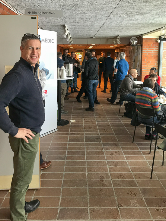
David O’Carroll was pleased with the introduction talk.
Yesterday the department’s new confocal and light sheet microscope, the Leica TCS SP8 DLS, was inaugurated at a highly successful launch event held in the Biology A and B buildings.
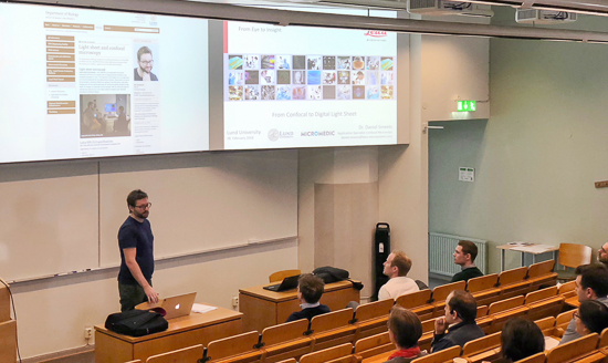
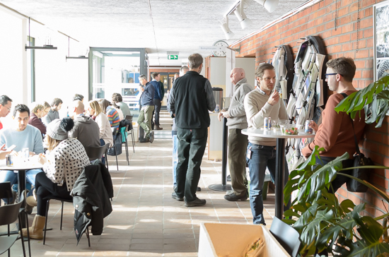
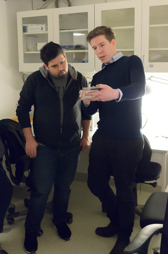
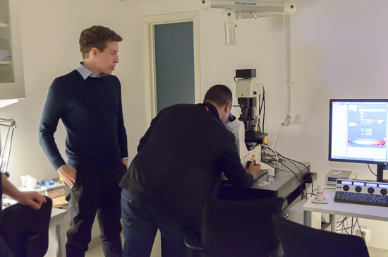
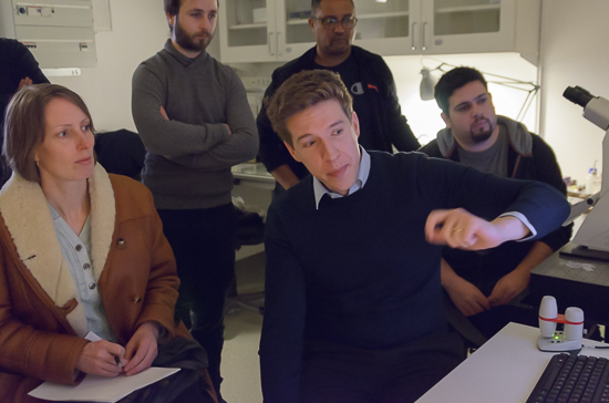
Despite the difficult morning weather, almost 50 people attended the morning presentation given by Micromedic and the Leica Microsystems’ application team. After lunch many of the participants were then able to see the new instrument at close hand, with demonstrations of sample preparation and the digital light-sheet imaging capability of the microscope, as well as an exhibition of Leica stereo and routine microscopes held by Micromedic.
Ola Gustafsson showed the updated website among other things.
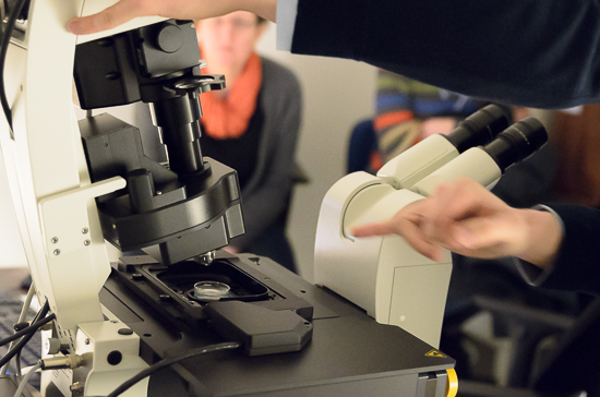
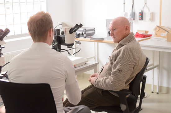
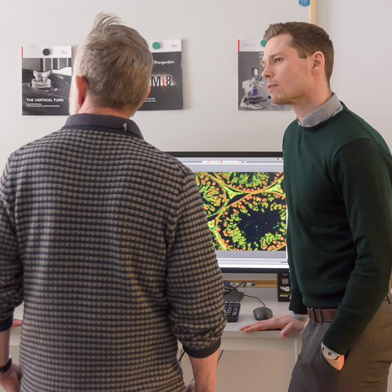
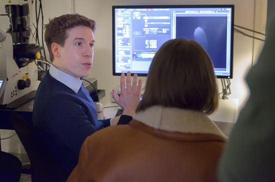
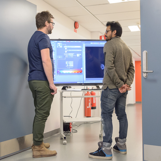
Lunch break after the morning presentations.A sample being placed into the light path ready for light sheet imaging.Applications specialist Daniel Smeets from Leica demonstrating how to prepare sample for light sheet imaging.David O’Carroll and Ola Gustafsson looking on during the light sheet imaging demonstration.Rolf Elofsson checking out some of the new stereomicroscopes being demonstrated by Micromedic.Rickard Linnskog from Micromedic with the Leica stereomicroscope system.Applications specialist Daniel Smeets with the department’s new Leica SP8 DLS system.The SP8 DLS was very popular during the light sheet imaging demos, with over 30 people seeing the system first hand over 4 sessions, and getting to watch Daniel Smeets building a 3D image stack with 70 slices in just 20 seconds from a GFP expressing Drosophila embryo.Applications specialist Daniel Smeets demonstrating the Leica software.With the light sheet demonstrations packed, there was still room in the corridor for people to see the DLS system mirrored on the big screen, or chat about their needs and book a session for checking out their own samples with Ola Gustafsson.